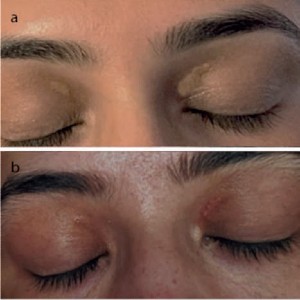
https://doi.org/10.4081/ltj.2023.317 Endolesional ablation of xanthelasma using microfiber optic laser delivery PDF Vol. 30 No. 2 (2023) Submitted: 28 May 2023 Accepted: 23 August 2023 Published: 12 October 2023 Xanthelasma, laser, microfiber, adipose, palpebrae, periorbital Abstract Views: 2249 PDF: 78 Publisher's note All claims expressed in this article are solely those of the authors and do not necessarily represent those of their affiliated organizations, or those of the publisher, the editors and the reviewers. Any product that may be evaluated in this article or claim that may be made by its manufacturer is not guaranteed or endorsed by the publisher. Authors Ernest A. Azzopardi info@skinsurgeon.eu Department of Surgery, Division of Interventional Sciences, University College London, London, United Kingdom; Skinsurgeon@ELms Clinic, Triq il Pirinej, Malta; Consultant Surgeon in Private Practice, The London Welbeck Hospital, Marylebone, London; Department of Anatomy, Faculty of Medicine and Surgery, University of Malta Medical School, Malta. Sarah Refalo Azzopardi Department of Ophthalmology, Mater Dei Hospital, United Kingdom. Matteo Tretti Clementoni Laserplast Srl Stp, Milan, United Kingdom. Dean Edward Boyce Welsh Centre for Plastic Surgery, Morriston Hospital, Abertawe, Wales, United Kingdom. Hazim Sadideen Welsh Centre for Plastic Surgery, Morriston Hospital, Abertawe, Wales, United Kingdom. Ash Mosahebi Department of Surgery, Division of Interventional Sciences, University College London, London, United Kingdom. Abstract Xanthelasma palpebrarum is the most common type of cutaneous xanthoma and is often a cause of psychological distress and aesthetic dissatisfaction. The extent, depth, or background skin type, intolerance to downtime, or cost, may restrict the treatment options, or contribute to a recurrence rate of up to 60%. 1470 nm microfiber laser is a recent clinical innovation that allows highly targeted delivery of Laser to deeper tissues through fibers as small as 150 μm in diameter, targeting fat and/or water chromophores. We report a retrospective data series on five patients (10 eyelids) treated with intralesional microfiber laser, where other treatment methods were inappropriate, contraindicated, or declined. Single-use tip firing microfibers (150-300 μm), were introduced into lesions under tactile and visible indicator light guidance (1-2 W; 250-500 Hz, LEED 1-2 Jcm–2, 1470 nm ). Results were followed up with before/after photography. The pain was measured using a prevalidated 1-10 Likert scale. Patients were followed up by remote consultation up to one year post-treatment. Xanthelasma size was (7 mm ± 4 mm, mean ±SD). The average time to complete resolution was 12±2.4 weeks ( All patients were normolipidemic pre-treatment. Sessions needed were 1.2±0.4 (mean ±SD). Maximum discomfort on a 1-10 Likert scale was 3±1/10 (mean ±SD), at eight weeks’ follow-up. No recurrences were reported up to 1 year’s follow-up. No patients had visible scarring. Most importantly, all patients reported minimal downtime and could continue normally with activities of daily life. 1470 nm microfiber laser is a promising method for the management of palpebral xanthelasma: within this case series was safe and effective in experienced hands. Further, larger studies are in hand to assess follow-up long-term outcomes and patient satisfaction. Metrics Dimensions Altmetric PlumX Metrics Downloads Download data is not yet available. Citations References Howick J, Phillips B, Ball C, Sackett D, Badenoch D. Oxford centre for evidence-based medicine levels of evidence. University of Oxford, Centre for Evidence-Based Medicine 2009. Rohrich RJ, Janis JE, Pownell PH. Xanthelasma palpebrarum: a review and current management principles. Plastic Reconstruct Surg 2002:110:1310-14. DOI: https://doi.org/10.1097/00006534-200210000-00016 Lee HY, Jin US, Minn KW, Park Y-O. Outcomes of surgical management of xanthelasma palpebrarum. Arch Plastic Surg 2013:40:380. DOI: https://doi.org/10.5999/aps.2013.40.4.380 Hawash AA, Ingrasci G, Nouri K, Yosipovitch G. Pruritus in keloid scars: mechanisms and treatments. Acta Dermato-Venereologica 2021:101:adv00582-adv82. DOI: https://doi.org/10.2340/00015555-3923 Esmat SM, Elramly AZ, Abdel Halim DM, et al. Fractional CO2 laser is an effective therapeutic modality for xanthelasma palpebrarum: a randomized clinical trial. Dermatol Surg 2014;40:1349-55. DOI: https://doi.org/10.1097/DSS.0000000000000172 Joshi A, Kale S, Chandel S, Pal DK. Likert scale: Explored and explained. Curr J Appl Sci Technol 2015;7:396-403. DOI: https://doi.org/10.9734/BJAST/2015/14975 Nahas TR, Marques JC, Nicoletti A, et al. Treatment of eyelid xanthelasma with 70% trichloroacetic acid. Ophthalm Plastic Reconstruct Surg 2009:25:280-83. DOI: https://doi.org/10.1097/IOP.0b013e3181aa9a1f Katz TM, Goldberg LH, Friedman PM. Fractional photothermolysis: a new therapeutic modality for xanthelasma. Arch Dermatol 2009:145:1091-94. DOI: https://doi.org/10.1001/archdermatol.2009.234 Mittelviefhaus H, Kreusser C, Böhringer D, Auw-Hädrich C. Die oft unterschätzte Tiefenausdehnung von Xanthelasmen – eine histologische Studie. Klinische Monatsblätter für Augenheilkunde 2011:228:14-18. DOI: https://doi.org/10.1055/s-0029-1245829 Laftah Z, Al-Niaimi F. Xanthelasma: An update on treatment modalities. J Cutaneous Aesthetic Surg 2018:11:1. DOI: https://doi.org/10.4103/JCAS.JCAS_56_17 Cannon P, Ajit R, Leatherbarrow B. Efficacy of trichloroacetic acid (95%) in the management of xanthelasma palpebrarum. Clin Exper Dermatol 2010:35:845-48. DOI: https://doi.org/10.1111/j.1365-2230.2010.03818.x Mourad B, Elgarhy LH, Ellakkawy HA, Elmahdy N. Assessment of efficacy and tolerability of different concentrations of trichloroacetic acid vs. carbon dioxide laser in treatment of xanthelasma palpebrarum. J Cosmetic Dermatol 2015:14:209-15. DOI: https://doi.org/10.1111/jocd.12148 Rossi AM, Perez MI. Treatment of hyperpigmentation. Facial Plastic Surgery Clinics 2011:19:313-24. DOI: https://doi.org/10.1016/j.fsc.2011.05.010 Park EJ, Youn SH, Cho EB, et al. Xanthelasma Palpebrarum Treatment with a 1,450‚Äênm‚ÄêDiode Laser. Dermatol Surg 2011:37:791-96. DOI: https://doi.org/10.1111/j.1524-4725.2011.01945..x Nguyen AH, Vaudreuil AM, Huerter CJ. Systematic review of laser therapy in xanthelasma palpebrarum. Int J Dermatol 2017:56:e47-e55. DOI: https://doi.org/10.1111/ijd.13534 Al-Kady NA, Hamdino M, Abdel Kawy FAW. Fractional CO2 laser versus trichloroacetic acid 50% for xanthelasma palpebrarum therapy. J Cosmet Dermatol 2021;20:763-768. DOI: https://doi.org/10.1111/jocd.13666 No D, McClaren M, Chotzen V, Kilmer SL. Sebaceous hyperplasia treated with a 1450-nm diode laser. Dermatol Surg 2004;30:382-4. DOI: https://doi.org/10.1111/j.1524-4725.2004.30105.x Echague AV, Astner S, Chen AA, Anderson RR. Multiple apocrine hidrocystoma of the face treated with a 1450-nm diode laser. Arch Dermatol 2005:141:1365-67. DOI: https://doi.org/10.1001/archderm.141.11.1365 Li K, Nicoli F, Xi WJ, et al. The 1470 nm diode laser with an intralesional fiber device: a proposed solution for the treatment of inflamed and infected keloids. Burns Trauma 2019;7:5. DOI: https://doi.org/10.1186/s41038-019-0143-6 Nilforoushzadeh MA, Fakhim T, Heidari-Kharaji M, et al. Efficacy evaluation of Endolift-based Subcision on acne scar treatment. J Cosmet Dermatol 2021;20:2579-2582. DOI: https://doi.org/10.1111/jocd.13876 Citation / Copyright How to Cite Azzopardi, E. A., Refalo Azzopardi, S., Tretti Clementoni, M., Boyce, D. E., Sadideen, H., & Mosahebi, A. (2023). Endolesional ablation of xanthelasma using microfiber optic laser delivery. Laser Therapy, 30(2). https://doi.org/10.4081/ltj.2023.317 More Citation Formats ACM ACS APA ABNT Chicago Harvard IEEE MLA Turabian Vancouver Download Citation Endnote/Zotero/Mendeley (RIS) BibTeX Copyright (c) 2023 the Author(s) This work is licensed under a Creative Commons Attribution-NonCommercial 4.0 International License. PAGEPress has chosen to apply the Creative Commons Attribution NonCommercial 4.0 International License (CC BY-NC 4.0) to all manuscripts to be published. Similar Articles Leonardo Longo, Roberto Dell'Avanzato, Diego Longo, ENDOLIFT® and multi-wavelength laser photobiomodulation: a randomized controlled trial study on 96 subjects, treating skin laxity of the lower third of the face , Laser Therapy: Vol. 29 No. 1 (2022) Luca Zattoni, Alessia Forlin, Real-life efficacy interstitial laser therapy treating laxity of the periorbital region , Laser Therapy: Vol. 30 No. 1 (2023) Diego Longo, Giulio Cherubini, Vanessa Mangè, Paolo Lippi, Leonardo Longo, Daniela Melchiorre, Maria Angela Bagni, Effects of laser therapy and Grimaldi’s muscle shortening maneuver on spasticity in central nervous system injuries , Laser Therapy: Vol. 29 No. 1 (2022) Sana Nadeem, Frequency-doubled neodymium yttrium aluminum garnet (Nd:YAG) laser trabeculoplasty as adjuvant therapy for open-angle glaucomas , Laser Therapy: Vol. 31 No. 1 (2024) Riccardo Lazzari, Carlo Fornaini, Leonardo Longo, Fractional treatment of rosacea by LBO 532 nm laser with “one shot” procedure: A preliminary study , Laser Therapy: Vol. 29 No. 2 (2022) Antonio Leccisotti, Stefania V. Fields, Giuseppe De Bartolo, Christian Crudale, Incidence and outcome of suction loss during corneal lenticule extraction with the femtosecond laser application CLEAR , Laser Therapy: Vol. 30 No. 1 (2023) Mahshid Saffarpour, Sajjad Gerami, Nastaran Parviz, Nasim Chiniforush, Evaluating the effectiveness of two wavelengths of 810 and 980 nm Diode laser with two different beam profiles on tooth discoloration in bleaching: an experimental study , Laser Therapy: Vol. 31 No. 1 (2024) Aizhan Issatayeva, Carlo Fornaini, Matteo Masino, Annamaria Cucinotta, Potential role of Er:YAG laser and fluoride in the dental enamel remineralization: a Raman spectroscopy preliminary ex vivo study , Laser Therapy: Vol. 29 No. 1 (2022) Antonio Leccisotti, Stefania V. Fields, Giuseppe De Bartolo, Christian Crudale, Effective optical zone after corneal lenticule extraction with the CLEAR femtosecond laser application , Laser Therapy: Vol. 30 No. 2 (2023) Leonardo Longo, Good news from China! , Laser Therapy: Vol. 31 No. 2 (2024) 1 2 3 4 5 > >> You may also start an advanced similarity search for this article.